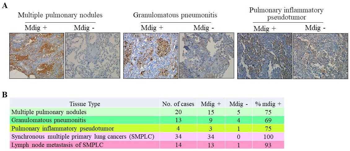
Fig. 6. Evaluation of mdig expression in pathological human pulmonary tissues. (A) Biopsies consisting of lesions harboring multiple pulmonary nodules, granulomatous pneumonitis and pulmonary inflammatory pseudotumor were processed for paraffin embedding, sectioning and staining for mdig protein. Samples were found to be positive and negative for mdig expression. Cellular localization of mdig was predominately nuclear, membranous and cytoplasmic in conjunction with nuclear staining. Multiple pulmonary nodules, n=20 cases, granulomatous pneumonitis, n=13 cases, pulmonary inflammatory pseudotumor, n=4 cases. (B) Summary of mdig expression among the indicated number of cases with non-cancerous lung diseases. For comparison, the mdig expression rate in SMPLC along with the metastatic lymph nodes was also included.